
Combining Multiple Z-Axis Images for Clearer Microscopic Observation with AxisPro
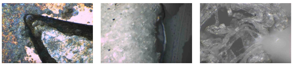
Microscopic samples are rarely flat. Surfaces can be uneven, and features often sit at different heights. Under a microscope, only part of such a sample is sharp at any one time. The rest drifts out of focus.
This comes down to depth of field, the range along the optical axis over which a sample appears acceptably sharp. Depth of field generally becomes shallower as magnification increases. A single image may therefore not keep the whole area of interest in focus.
One practical answer is to capture multiple images at different Z-axis positions and combine them. This approach is often called focus stacking, and MicroSupport describes it as focus synthesis. MicroSupport documents it with the AxisPro: it captures images at multiple Z-axis positions, and optional image-composition software combines them into a single view.
Why One Microscopic Image May Not Show the Entire Sample Clearly
In a single image, only a limited depth range is sharply focused. Vertically extended features, or an uneven surface, fall at different focal positions, so some regions look sharp while others look blurred.
Changing the focus position brings different parts of the sample into sharp focus. Capturing images at several focus positions therefore records every region of the sample at least once in focus.
Image composition combines information from these different focal positions. It does not physically increase the optical depth of field of the microscope.
Capturing Multiple Images Along the Z-Axis
Controlled Z-Axis Movement
Moving along the Z-axis changes the focus position. Each position corresponds to a different focal plane, which brings a different depth of the sample into focus. Imaging conditions need to stay stable throughout the sequence so that the images can be combined cleanly.
Barnett describes the AxisPro as incorporating a motorized XYZ stage, with all motion controlled through software.
Sequential Image Acquisition
The basic workflow is:
- Position the sample under the microscope.
- Identify the area of interest.
- Establish the Z-axis focus range required.
- Capture images at successive Z-axis positions.
- Pass the image sequence to the optional image-composition software.
- Combine the images to produce the composite view.
The number of images and the Z-axis step size depend on the sample and the system configuration. MicroSupport does not specify fixed values for this application.
AxisPro Image Composition Workflow
The process runs in five stages:
Sample Positioning → Z-Axis Movement → Image Capture → Image Composition → Final Observation
- Sample positioning: Position the sample and identify the area to be observed.
- Z-axis movement: Move through the required focal range along the Z-axis.
- Image capture: Capture multiple images corresponding to different focal positions.
- Image composition: Use the optional software to combine the captured images. The composite uses information from the different focal planes to show a greater portion of the sample in focus.
- Final observation: Use the resulting composite image for clearer observation and documentation of the sample.
What Z-Axis Image Composition Provides
Combining images from multiple focal planes can produce a composite with a greater apparent in-focus region than any individual image. That can help with visualizing samples that have uneven surfaces or features at different heights.
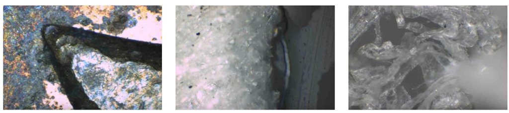
Three points keep expectations accurate:
- The final image is computationally composed from the captured image sequence.
- It adds no new optical information. It can only combine detail the microscope actually captured.
- It is not a 3D model or depth map. MicroSupport describes the sample in this application as three-dimensional, meaning a physical sample with height variation. The composite itself is a flat image for observation and documentation.
Role of AxisPro in the Image-Capture Process
The AxisPro provides controlled positioning for the image-capture process. Barnett describes it as a benchtop micromanipulator incorporating a zoom microscope, a motorized stage, and two motorized arms, with motion controlled through software. Controlled Z-axis positioning supports repeatable capture of images at different focal positions.
Each part of the setup has its own role:
| Component | Role |
| AxisPro | Provides controlled positioning and image acquisition support |
| Microscope | Provides the optical image (on the AxisPro, the integrated zoom microscope) |
| Image-composition software | Combines the multiple images into the composite result |
Watch the video demonstration to see how AxisPro captures multiple images at different Z-axis positions and combines them using optional image-composition software.
Applications of Z-Axis Image Composition
Observing a larger portion of a sample in focus may be useful in areas such as:
- Microscopic inspection of uneven surfaces
- Observation of small components
- Electronic component inspection
- Microscopic sample documentation
- Surface-feature observation
- Research and laboratory microscopy
- Observation of samples with features at different heights
These are potential application areas. They are not a claim that the manufacturer has demonstrated each one.
Factors That Can Affect Image Composition
Several factors influence how well the composite turns out:
- Sample height variation: The more the sample’s height varies compared with the depth of field, the more focal coverage is needed.
- Microscope magnification: Higher magnification generally narrows the depth of field.
- Depth of field: It determines how much of the sample is sharp in each image.
- Z-axis travel range: The range must cover the sample’s height variation.
- Focus-step selection: Steps that are too large can leave gaps in focal coverage, while very small steps may produce more images than needed.
- Image quality: The composite can only draw on the quality of the source images.
- Sample stability: Movement during the sequence can degrade the result.
- Illumination consistency: Brightness changes between images can affect the composite.
- Optical alignment: Consistent alignment helps the images combine cleanly.
- Image-processing and software settings: Software settings can influence the final image.
Suitable parameter values depend on the specific AxisPro configuration and the sample, so none are given here.
Best Practices for Z-Axis Image Acquisition
- Secure the sample first: Fix it before starting image acquisition.
- Select suitable magnification: Match it to the area and detail needed.
- Identify the focal limits: Determine the upper and lower focal positions required.
- Keep illumination consistent: Maintain it throughout the capture.
- Prevent sample movement: Keep the sample still for the whole sequence.
- Choose appropriate Z-axis increments: Match them to the sample and optical setup.
- Cover the area of interest: Capture sufficient focal coverage across it.
- Review the individual images: Check them before composition when necessary.
- Use the supported software configuration: Follow the manufacturer’s configuration for image composition.
Key Benefits
- A larger apparent in-focus region in the final composite image
- Less dependence on a single focal plane
- Clearer observation of uneven microscopic features
- Controlled Z-axis image acquisition
- Repeatable focus positioning
- Motorized microscope positioning combined with software-based image composition
- Better visual documentation of microscopic samples
Samples that extend across multiple focal planes cannot be fully sharp in a single image. Capturing images at different Z-axis positions records each part of the sample in focus, and optional image-composition software combines those images into one composite view. The AxisPro provides the controlled positioning for image acquisition, and the software performs the composition. Together they can support clearer microscopic observation and documentation of samples with varying surface heights, without claiming a 3D reconstruction.
Barnett Technical Services provides AxisPro microscope-integrated micromanipulation systems and MicroSupport solutions for precision microscopic observation, sample handling, and image-based workflows. Contact us to discuss your microscopy and micro-analysis requirements.