An Introduction to Coherent Anti-Stokes Raman Microscopy
In the ever-evolving field of optical microscopy, Coherent Anti-Stokes Raman Spectroscopy (CARS) has emerged as a powerful, label-free imaging technique for probing the molecular composition of biological and chemical samples. Among the family of Coherent Raman Scattering techniques, CARS stands out for its exceptional signal strength, high spatial resolution, and ability to perform real-time, non-destructive chemical imaging.
But what makes CARS microscopy so transformative? Let’s explore the fundamentals and benefits of this advanced imaging method.
The Foundations: From Linear to Nonlinear Spectroscopy
Traditional Raman spectroscopy has long enabled scientists to analyse molecular vibrations and chemical structures by observing spontaneous inelastic scattering of light. However, the inherently weak signals and long acquisition times limit its use in dynamic or live systems.
Nonlinear spectroscopy overcomes these limitations by introducing multiphoton interactions. Specifically, third-order nonlinear optical processes are used to generate stronger and more coherent signals. These processes occur when high-intensity laser beams interact with materials in a way that nonlinear polarization is induced, leading to new frequencies of scattered light.
What is CARS Microscopy?
CARS is a third-order nonlinear optical process that amplifies the vibrational signals of molecules through coherent interactions between laser beams and molecular bonds.
In CARS microscopy, three beams interact within a sample:
- A pump beam at frequency νₚ
- A Stokes beam at a lower frequency νₛ
- A probe beam, usually at the same frequency as the pump (νₚ = νₚᵣₒᵦₑ)
When the difference between the pump and Stokes frequencies matches a specific vibrational mode of a molecule (νₚ – νₛ = νᵥᵢᵦ), a coherent vibration is induced. This leads to the generation of an anti-Stokes signal at a higher frequency νcars = 2νₚ – νₛ. Unlike spontaneous Raman scattering, the CARS signal is directional, coherent, and several orders of magnitude stronger, enabling faster imaging with higher resolution and less sample damage.
Advantages of CARS Microscopy
CARS offers numerous benefits over traditional Raman and fluorescence microscopy:
1. Ultra-High Sensitivity
CARS signals can be 104-105 times stronger than those from spontaneous Raman scattering. This allows for fast signal acquisition enabling real-time imaging.
2. Label-Free Imaging
Unlike fluorescence microscopy, CARS does not require labeling or staining, making it ideal for studying live cells, tissues, and dynamic chemical processes.
3. Optical Sectioning and 3D Imaging
The CARS signal originates only from the tight focal volume where the beams overlap, allowing precise spatial localization and high-resolution 3D sectioning.
4. Suppression of Fluorescence Background
Since the anti-Stokes signal lies at a higher energy than the excitation sources, it can be easily separatedfrom fluorescence and other background signals using optical filters.
5. Quantitative Capability
CARS intensity depends quadratically on the concentration of resonant molecules, making it suitable for quantitative chemical mapping when properly calibrated.
Technical Insights: Resonance and Signal Generation
CARS is most efficient when the laser frequency difference matches a molecule’s vibrational frequency. The signal intensity scales as:
I₍CARS₎ ∝ |χ⁽³⁾|² × Iₚᵤₘₚ² × Iₛₜₒₖₑₛ
where:
- χ(3) represents the third-order susceptibility tensor.
- Ipump> and Istokes are the intensities of the pump and Stokes beams.
The χ(3) term captures both resonant and non-resonant contributions. The resonant term corresponds to the targeted molecular vibration, while the non-resonant background arises from off-resonance interactions. This can sometimes obscure the signal but can be mitigated through polarization-sensitive detection (P-CARS) or epi-detection (E-CARS).
Forward vs. Backward Detection
- F-CARS (Forward CARS): High signal strength but may suffer from non-resonant background.
- E-CARS (Epi CARS): Weaker signal, but with minimal non-resonant interference, ideal for imaging small or sub-wavelength-sized structures.
Applications of CARS Microscopy
CARS microscopy has proven transformative across multiple disciplines:
- Cell Biology: Visualize lipids, proteins, and organelles in live cells without dyes.
- Neuroscience: Map myelin sheaths and monitor brain tissue health.
- Pharmaceuticals: Analyze drug distribution in tissues and tablets.
- Chemical Engineering: Study combustion processes and catalyst surfaces.
- Material Science: Examine polymers, composites, and nanomaterials.
Comparison Table: CARS vs. Spontaneous Raman vs. SRS
| Feature | CARS | Spontaneous Raman | SRS |
| Signal Strength | Very High (coherent) | Low (incoherent) | High (linear) |
| Label-Free | Yes | Yes | Yes |
| Non-resonant Background | Yes (unless suppressed) | No | No |
| Fluorescence Interference | None | Can be severe | None |
| Imaging Speed | Fast | Slow | Fast |
| Spectral Info | Moderate (unless multiplexed) | Rich | Limited (unless swept) |
Final Thoughts on CARS Microscopy
Coherent Raman Scattering Microscopy, particularly CARS, offers a powerful toolkit for non-invasive, high-speed, and high-resolution imaging at the molecular level. By bridging the gap between spectroscopy and microscopy, CARS enables scientists to visualize chemical landscapes with unprecedented clarity.
As instrumentation advances and signal processing improves, CARS is poised to play a leading role in fields ranging from biomedical diagnosticsto industrial quality control.
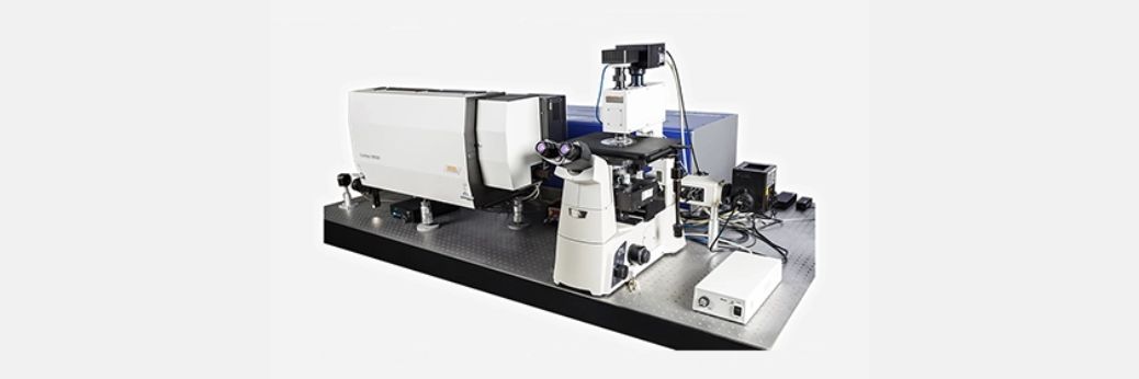
At Barnett Technical Services, we offer the Confotec® CARS system, which combines a CARS scanning microscope, a Raman/luminescent scanning confocal microscope, and a conventional scanning confocal laser microscope in a single integrated platform—delivering unmatched versatility for advanced molecular imaging.