Confocal Raman Microscopy: Principles, Capabilities, and Real-World Applications
In modern materials science, nanotechnology, and life sciences, the ability to chemically image microscopic structures with high precision is no longer a luxury—it’s a necessity. Confocal Raman Microscopy has emerged as a powerful, non-destructive analytical technique that meets this need by combining Raman spectroscopy with the spatial resolution of confocal optical microscopy.
But what exactly is Confocal Raman Microscopy, and how does it work?
What Is Confocal Raman Microscopy?
Confocal Raman Microscopy integrates the molecular specificity of Raman spectroscopy with the high-resolution imaging capabilities of a confocal microscope. It enables 2D and 3D chemical imaging of samples at sub-micron resolution without requiring sample preparation.
This technique is capable of identifying and mapping the chemical composition of a sample in three dimensions—making it ideal for analyzing multilayer films, polymers, semiconductors, pharmaceuticals, and biological materials.
How Does Confocal Raman Microscopy Work?
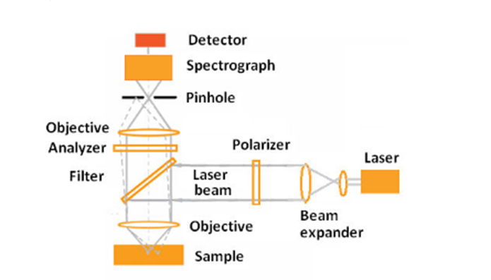
The core of this technology lies in combining several key optical components:
1. Laser Excitation : A monochromatic laser (e.g., 532 nm, 633 nm, or 785 nm) is used to illuminate a precise spot on the sample. The high-intensity laser light enhances the inherently weak Raman scattering signals.
2. Objective Lens: The objective lens focuses the laser onto the sample and collects the back-scattered light. Its numerical aperture (NA) plays a vital role in determining lateral and axial resolution. For example, with a 514.5 nm laser and an NA of 0.9, lateral resolution can reach ~360 nm.
3. Rejection Filters : To isolate the Raman signal, Rayleigh scattered light (which carries no chemical information) is removed using edge or notch filters. SOL Instruments, for example, offers BragGrate notch filters with bandwidths <10 cm⁻¹, allowing ultra-low-frequency Raman analysis.
4. Confocal Pinhole : A confocal pinhole eliminates out-of-focus light, ensuring only in-focus Raman signals are detected. This spatial filtering improves both image contrast and axial (Z-axis) resolution, which is critical for 3D Raman mapping.
5. Spectrograph and Detectors: The Raman scattered light is dispersed by a diffraction grating inside a spectrograph and captured by a CCD or EMCCD detector. High-resolution gratings (e.g., 1800 lines/mm or echelle gratings) allow the detection of subtle chemical differences, while EMCCDs enable fast, sensitive data collection.
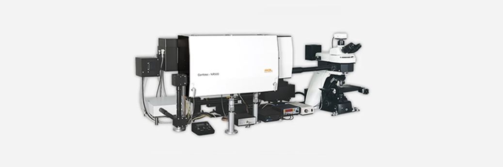
Advanced Capabilities: Confotec® Raman Systems
SOL Instruments’ Confotec® series, such as the NR500, exemplify state-of-the-art Confocal Raman systems. These instruments can be configured with up to five laser lines and ultra-low frequency Raman filters, enabling the detection of Stokes and anti-Stokes signals down to 10 cm⁻¹—ideal for probing phonon modes, carbon nanotubes, and 2D materials like graphene. They also feature:
- 3D scanning capabilities
- Temperature-controlled stages (from -196°C to 1000°C)
- High spectral resolution (FWHM as low as 0.017 nm with echelle gratings)
- Volume Bragg Gratings (VBGs) for stable, high-throughput filtering
Applications of Confocal Raman Microscopy
The versatility of Confocal Raman Microscopy allows it to be used in an impressive range of disciplines:
Materials Science
- Stress mapping in silicon wafers
- Polymorphic analysis of pharmaceuticals
Nanotechnology
- Characterization of carbon nanotubes (RBM, D-band, G-band)
- Graphene layer identification via C-band
Life Sciences
- Protein structure analysis (e.g., keratin in nails)
- Cell and tissue biochemistry
- Degree of crystallinity in cellulose
Semiconductor and Electronics
- Analysis of Li-ion battery degradation
- Mapping semiconductor heterostructures (Si-Ge, GaN, QDs)
Forensics & Document Analysis
- Ink discrimination
- Sequence of handwritten layers
- Pigment analysis through packaging
Advantages of Confocal Raman Microscopy
- Label-Free and Non-Destructive
- High Spatial and Spectral Resolution
- 3D Chemical Mapping
- Minimal Sample Prep
- Simultaneous Imaging and Spectroscopy
Key Insights from Confocal Raman Microscopy
Confocal Raman Microscopy stands at the intersection of chemical insight and optical precision. Whether you’re investigating strain in semiconductors, crystal structures, or molecular compositions deep within a material, this technique offers a depth of information unmatched by traditional spectroscopy or microscopy alone.
If you’re looking for a system that combines sensitivity, resolution, and flexibility—SOL Instruments’ Confotec® series should be on your radar.
Looking to Upgrade Your Lab’s Imaging Capabilities?
Contact Barnett Technical Services to learn more about Confocal Raman Microscopes and how they can be configured for your research or industrial application.