Coherent Anti-Stokes Raman Scattering (CARS) microscopy permits the acquisition of vibrational spectra for many samples where signa intensity, fluorescence, or other properties of the sample make it difficult to obtain structural information. This technique is especially valuable for biological samples which were viewed using stains and dyes for differentiation. However, this method had various drawbacks, including the bleaching of dyes over time and potential cytotoxicity, which could harm the samples or interfere with results. A CARS microscope offers simultaneous multifunctional and high-speed imaging of samples using 3D scanning laser spectroscopy. At Barnett Technical Services, we offer the Confotec® CARS system that combines a CARS scanning microscope, a Raman/luminescent scanning confocal microscope, and a conventional scanning confocal laser microscope in a single system.
CARS Microscope
Products
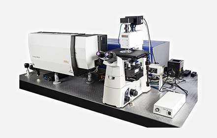
The Confotec® CARS System is the multifunctional multi-channel 3D scanning laser microsco..
Read More..Key Features of CARS Microscopes
Here are some key features of CARS microscopes- High-Resolution and Label-Free Imaging: CARS signal is proportional to the squared molecule concentration, and hence applicable for quantitative measurements of chemical substance concentration in a sample. It does not require chemical labeling, and offers high speed imaging and resolution.
- Real-Time and Live Cell Imaging: The sample cells remain fresh and are not damaged as dyes and labeling are not used. Scientists get to study live cells in real time without photodamage.
- Compatibility with Other Imaging Modalities: In two photon fluorescence, two photons are absorbed at a low energy state, after which one of its electrons temporarily converts to a higher energy state and emits a single excited photon. This energy level of the emitted excited photon is higher than that of the absorbed photons. Second harmonic generation (SHG) comprises a laser light focused on a sample to generate frequency-doubled light. This means two photons of the same wavelength are annihilated generating a photon of half that wavelength. CARS microscopy is compatible with other imaging modalities such as confocal microscopy, two photon fluorescence, second harmonic generation, and so on.
How Does a CARS Microscope Work?
The Confotec® CARS system is a>multi-channel 3D scanning laser microscope-spectrometer that offers confocal Raman imaging, confocal fluorescence imaging, and confocal imaging in the reflected laser radiation, high-contrast imaging in the transmitted laser radiation, and visualization of a surface profile through second harmonics generation signal. Here are some pointers on how CARS microscopy works.
- CARS microscopy generates more intensive and directed signals than Raman microscopy. It enables detection of signals in a fluorescence-free spectral range, unlike conventional microscopy that is still widely used.
- CARS microscopy is based on the Raman effect, wherein incident light scatters interacts with a sample. Some scattered photons may have a wavelength different from that of the incident light due to the excitation of the object molecules at high energy levels.
- Raman spectroscopy often requires a long integration time to collect any real-time data. As a non-linear advanced spectroscopy technique, CARS overcomes this issue of imaging speed making the collection of real-time data possible.
- In CARS microscopy, two beams of light are used to simultaneously excite the sample, wherein one beam is referred to as the pump beam and the other is the stokes beam. When the frequency difference between the two beams of light corresponds to the vibrational frequency of the sample, four simultaneous coherent Raman processes occur.
- This non-invasive method offers a high resolution of samples such as of biomolecules without labelling, and using non-confocal pinholes. Hence, the sample remains untouched and unaffected.
- CARS microscopy also performs 3D layer-by-layer scanning with minimal influence of neighboring layers on measurement results.
Applications of CARS Microscopy
Here are some application areas of CARS microscopy.- Biomedical research: This is the most common application of CARS microscopy as live biological samples can be used to study lipid and protein structures, neuroscience, cancer cells, live tissues, and so on.
- Materials Science: CARS microscopy enables non-destructive testing of polymers, ceramics, and other materials and helps boost the research on nanostructures.
- Environmental Science and Other Emerging Fields: The main segments in this area include chemical imaging in environmental analysis and forensics.