Confotec® MR series Raman Microscopes
Raman microscopes of Confotec® MR series
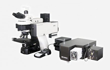
Compact Raman confocal microscopes-spectrometers of MR series from SOL instruments – models Confotec® MR350, Confotec® MR520 and Confotec® MR750 – are intended for micro spectroscopic measurements with the high-end system capabilities.
All models have a rigid design that does not require adjustments and have both high sensitivity and high spatial resolution. Spectral measurements are provided by a highly automated 350, 520 or 750 mm focal length Imaging monochromator-spectrograph with a motorized 4-position grating turret.
We offer 5 configurations for each model of Confotec® MR series microscopes so that you can select the optimal assembly for your industrial or scientific task.
- Modular design: the main Raman optical unit with a built-in laser is attached to the microscope, and the fiber coupled spectrograph can be arbitrarily located on a desktop or optical table.
- Rigid design provides high thermal and temporal stability of spectral measurements, no pre-adjustment is needed.
- Nondestructive analysis, no special requirements for sample preparations.
- Two automatic switchable lasers with different wavelengths allow to measure Raman spectra of various samples without influence of fluorescence background.
- Highly automated 350, 520 or 750 mm focal length Imaging monochromator-spectrograph with a motorized four-position grating turret provides spectral measurements.
- Only some sample areas are located in focus due to limited confocal depth of field. A high-speed reflected unit allows you to quickly find a sample focus areas and use them for Raman measurements. This provides a significant gain in time and allows you to immediately use a high numerical aperture objective to record high spatial resolution Raman maps.
- High speed simultaneous Raman and Rayleigh sample imaging using PMT detectors and a XY galvano-mirror scanner.
- High speed and high spatial resolution ultra-wide panoramic imaging (Raman and Rayleigh scattering) using high speed PMTs, XY galvano-mirrors and an automated microscope stage.
- 2D / 3D Raman imaging of a sample surface using XY galvano-mirror / Z-piezo scanners.
- Automatic spectral calibration.